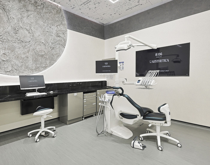
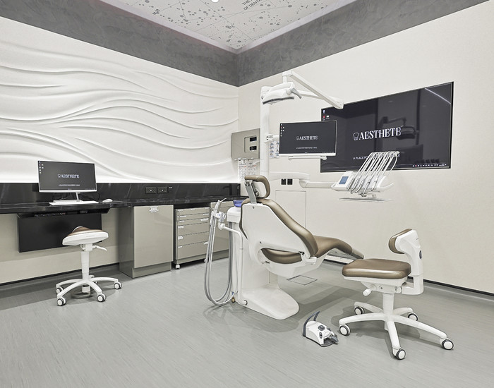
Tooth enamel erosion is a non-cariogenic disease that deteriorates the aesthetics of a smile and increases the risk of caries and other dental diseases. Statistics indicate that it is diagnosed in approximately one in four adults. AESTHETE Dental Clinic offers services for treating enamel erosion and its complications.
How we can be
useful to you:
Veneers Without Grinding
Pediatric dentistry
Correction of bite
Implantation and crowns
Main Causes of Tooth Enamel Erosion
Modern medicine does not fully understand enamel erosion, and some aspects of its onset are still under-researched. However, there are known causes that definitely contribute to the disease, such as:
- Damage due to strong mechanical impacts on the teeth – usually occurring during brushing when using too hard a brush, toothpaste, or powder with a high content of abrasive particles, but can also result from bruxism, involuntary jaw clenching;
- Chemical damage – caused by consuming foods and drinks high in acids or sugars: citrus fruits, pickled foods, sodas, energy drinks, sweet tea or coffee;
- Frequent intake of ascorbic acid supplements;
- Gastrointestinal diseases accompanied by high acidity levels;
- Reduced levels of ionized calcium and magnesium in the blood;
- Endocrine disorders, thyroid diseases – saliva becomes viscous and has a destructive effect on the hard tissues of the tooth;
- Uneven load on the teeth while chewing – due to the habit of chewing on one side, the absence of molars and premolars, or bite defects;
- Working in hazardous environments, such as those with metal dust and acid fumes in the air;
- Unfavorable environmental conditions;
- Age-related changes in the body.
Intense brushing and lemon juice treatment for whitening at home can also be triggering factors. In fact, those at risk are people who pay excessive attention to dental hygiene, as well as those who drink a lot of sodas, consume excessive amounts of fresh vegetables and fruits, and drink a lot of water with lemon.
Symptoms of Tooth Erosion
This pathology is more common in people aged 30-40 years. It develops gradually, sometimes taking 10-15 years from the onset of the first signs to the exposure of the dentin. A characteristic feature of the disease is that visible changes may not appear immediately. Often, enamel destruction occurs slowly and can only be noticed during a dental examination using magnifying devices. Therefore, it is important to visit the dentist at least twice a year to detect the early signs of the disease, allowing for easier treatment.
Visible manifestations of the disease resemble caries but have significant differences. Affected areas are round or oval, with a smooth, hard, shiny bottom. Initially, the color of the enamel remains unchanged, just losing its luster, but over time, the cavities turn yellow or brown. Usually, erosion affects at least two symmetrical teeth, most often the upper incisors, and less frequently the canines and premolars of both jaws. Lower jaw incisors and molars are generally not affected by this disease.
As the disease progresses, tooth sensitivity increases. They react painfully to touch during brushing and to hot, cold, or acidic foods.
Pathogenesis of Tooth Erosion
Tooth enamel is the hardest tissue in the human body. It is a thin transparent shell that covers dentin and pulp, serving a protective function. The enamel layer withstands mechanical and chemical impacts and temperature changes but can crack and chip. Unlike bone tissue, it does not regenerate.
An additional protective layer called the pellicle forms on the enamel surface after eating, composed of components from saliva. The pellicle protects the hard tissues of the teeth from acid impact but also contributes to bacterial plaque formation. Erosion starts when acids, metabolic disturbances, or strong mechanical impact remove the pellicle.
The next stage is the gradual demineralization of the enamel, losing substances that give it strength. At the neck of the tooth, where enamel is particularly thin, characteristic defects of a saucer-like shape with a smooth shiny bottom appear.
The affected area grows in width and depth. When enamel thins, the disease spreads to the dentin, forming concave defects because dentin is less strong.
Gum fluid partially slows down erosion by neutralizing acidic environments and replenishing mineral deficiencies in the enamel. However, without proper treatment, if aggressive factors continue to affect the teeth, the disease will progress.
-
Tooth Erosion Treatment cost
-
Tooth Erosion Treatment
from 860 AED
Classification and Stages of Tooth Erosion
There are two forms of the disease – active and stabilized. They are often considered as phases that follow one another. Signs of the active form include:
- Rapid destruction of the enamel layer, increasing erosion focus every 6-8 weeks;
- Sharp increase in tooth sensitivity, prompting patients to visit a dental clinic;
- The surface of the teeth becomes matte when dried, forming a plaque that is difficult to remove.
In the stabilized form:
- The disease progression slows down;
- Increased sensitivity, discomfort, and pain are almost absent;
- The affected area stabilizes, potentially remaining unchanged for 9-11 months.
Three stages of erosion development are also distinguished.
- Stage I – initial or superficial. At this stage, only the upper enamel layer is affected, covering no more than 1/3 of the tooth surface. The patient does not feel pain or discomfort when brushing or eating.
- Stage II – middle or transitional. The affected area increases, covering 1/3 to 2/3 of the tooth surface. The enamel layer is significantly damaged, down to the dentin, causing hypersensitivity and pain.
- Stage III – deep. Erosion affects the upper layer of the dentin, covering more than 2/3 of the tooth surface, with dark, highly visible spots.
Complications
Regardless of the erosion degree, the patient's quality of life deteriorates. Although there is no pain at the initial stage, the aesthetics of the smile worsen: dull spots appear on the tooth surface that resemble plaque but cannot be removed by brushing. Such spots indicate the onset of enamel destruction, making it softer and more sensitive to external factors.
As dentin becomes exposed, cosmetic defects become more noticeable. Yellow or brown spots appear, sharply contrasting with the light enamel. The disease more often affects the upper jaw's incisors, molars, and premolars, making the affected areas highly visible when smiling or talking.
Exposed dentin is sensitive to touch, causing pain while chewing. If the cause of the pain is not eliminated, gastrointestinal diseases may develop.
Dentin gradually deteriorates, involving the pulp, leading to other diseases such as pulpitis and periodontitis, potentially resulting in tooth loss.
Diagnosis of Tooth Erosion
Early signs of enamel layer destruction can only be noticed during a dental examination. If not detected early, the patient may visit the clinic with complaints of pain and discomfort, indicating that the disease has reached the second degree of severity.
The dentist listens to complaints and collects the medical history. Accurate responses help in diagnosing. At this stage, the specialist can identify the cause of erosion: for example, working in harsh, harmful conditions, a stomach disease causing acid reflux, or excessive citrus consumption.
The dentist then conducts an examination using a mirror and probe, identifying characteristic disease signs:
- Hard bottom, no pain when touched by the probe;
- Painful reaction to drying with air;
- Staining when exposed to iodine solution.
It is essential to confirm that it is erosion and not caries. Therefore, the dentist considers:
- The patient’s age – non-cariogenic enamel layer damage is typical for middle-aged people;
- Location of affected areas – usually symmetrical teeth, with defects on the most convex surface;
- Shape of the cavity – cup-shaped or saucer-shaped, with clear boundaries.
If enamel layer destruction is linked to endocrine disorders or gastrointestinal diseases, consultation with an endocrinologist or gastroenterologist is required to identify and eliminate the causes of tooth defects.
To make you healthy,
happy and successful!
will help you with this
How Tooth Enamel Erosion is Treated
Treatment must be comprehensive. The dentist needs to:
- Stop enamel layer destruction;
- Eliminate the disease-causing factor.
At the early stage, enamel condition can be stabilized with local remineralization using fluoride and calcium-containing preparations. The goal is to restore and strengthen the tooth’s protective layer. Applications are done daily for 15-20 days, followed by applying fluoride varnish to reduce sensitivity to temperature changes and acidic environments.
At the middle and severe stages, remineralization is also indicated, but it cannot fully resolve the problem or restore affected hard tissues. Therefore, restoration using composite materials or aesthetic defect elimination with crowns or veneers is necessary. AESTHETE dental clinic uses Magicneers technology to install veneers without tooth grinding.
The prognosis for erosion treatment is generally favorable, with tooth preservation. However, the earlier you seek medical attention, the easier it is to resolve the problem without crowns or veneers. If the disease severity reaches the third degree, tooth loss is possible.
Prevention
Unlike caries and other dental diseases, erosion development may be linked to excessive dental hygiene rather than its lack. Therefore, teeth should be brushed correctly. It is important to choose a brush with suitable bristle hardness and a non-destructive toothpaste. Avoid pressing too hard while brushing to prevent micro-scratches.
Other recommendations include:
- Avoid carbonated drinks, especially sweet ones;
- Drink juices with a straw to minimize contact with teeth, and if you can’t avoid soda, drink it through a straw too;
- Rinse your mouth after eating, preferably with antibacterial solutions rather than plain water;
- If rinsing isn’t possible, use chewing gum for no more than 5 minutes to clean teeth from small food particles;
- Eat fewer acidic foods, including fruits, vegetables, and dishes with vinegar;
- After meals, consume a piece of hard cheese to reduce oral cavity acidity.
Visit the dentist twice a year for preventive purposes.
AESTHETE dental clinic is located in Dubai (UAE), Bluewaters Island. We treat tooth erosion in patients of all ages using effective modern techniques. Our doctors will diagnose, make an accurate diagnosis, and provide the cost of services. They will develop a treatment plan and ensure the patient has no complications.
To make an appointment, contact the clinic administrators. They will answer your questions, help you choose a time for a dentist visit, and explain how to schedule a consultation.
Evaluate the amazing
results of our clients:
Literature sources
- Bartlett, D. W.; Smith, B. G. N.. Definition, classification and clinical assessment of attrition, erosion and abrasion of enamel and dentine. In: M. Addy, G. Embery, W. M. Edgar, R. Orchardson (eds.). Tooth wear and sensitivity - clinical advances in restorative dentistry . 1st ed., pp. 87–93. London: Martin Dunitz, 2000.
- Lussi, A.. Dental erosion; Clinical diagnosis and case history taking. European Journal of Oral Sciences , 2002, 104: 191–198.
- Grenby, T. H.. Methods of assessing erosion and erosive potential. European Journal of Oral Sciences , 1996, 104: 207–214.
Our clinics:
Clinic in Dubai
Resident part, Building 10
Clinic in Moscow
metro Chistye prudy
Clinic in Moscow (Barvikha)
Shopping center "Dream House" 3rd floor


_700x550_ac7.jpg)


